Last week saw me doing a long day down to the
Diamond light source to help with some sample preparation for an experiment next week that I am doing with
Liane Benning from Leeds. I had to get up at 5.30 in the morning and was back home by 11 in the evening; I think I'm still recovering.
The experiment next week will involve looking at thin slices through 2 mm diameter balls produced by earthworms.
 |
| 2mm diameter ball of calcium carbonate secreted by a Lumbricus terrestris earthworm |
The balls are made of small crystals of calcium carbon and the individual crystals are sometimes calcite, sometime aragonite, sometimes vaterite and sometimes amorphous calcium carbonate. These are all different forms of calcium carbonate, they vary in the arrangement of the calcium and carbonate molecules and we want to know what these different forms all occur together. The first step towards this aim is to map out the distribution of the different forms.
To do this we're going to do something called
FTIR (fourier transform infrared) mapping on
Beamline B22 at Diamond and to do that we need thin slices of the granules.
Just as there are many ways to skin a cat there are many ways to slice a granule. Last week "we" tried something called cryomicrotoming. We'd collected some fresh granules and kept them at -80 C so that they were preserved just as we had collected them. Then you simply attach them to a widget and slice them, rather like a salami slicer in the supermarket. It sounds easy but the slices are 5 - 10 micron thick (i.e. about one hundred times thinner than a finger nail) and the blade is in a small container into which liquid nitrogen is being pumped to keep the temperature low (about - 30 C) so it is rather fiddly. So fiddly that really the only person who was successful was the beamline scientist Katia Webhe who was there to give us a hand - many thanks Katia.
Katia Wehbe in action on the cryomicrotome, an admiring Mark Frogley looking on. Both Katia and Mark work on the beamline and help people like me get useful data. They are worth more than their weight in gold.
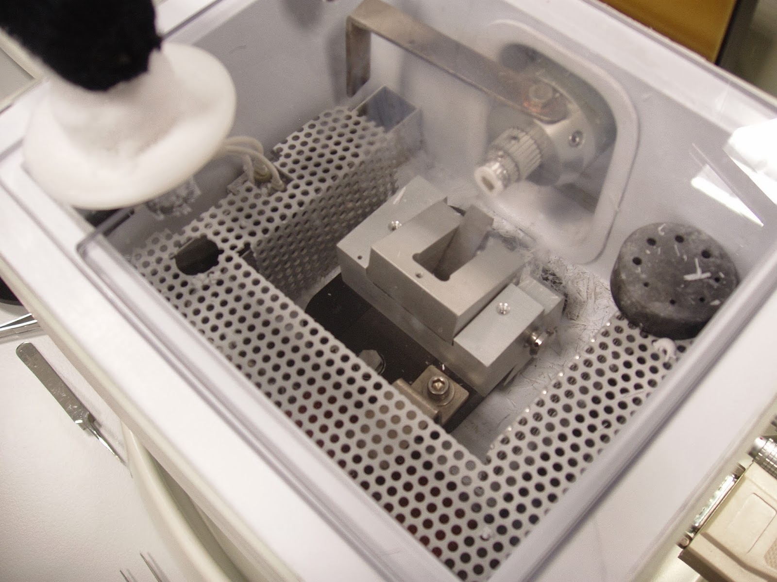
 |
| Inside the cryomicrotome. The white disk, top left is the liquid nitrogen outlet. Over to the right you can see a smaller white disk with a dark centre - that is our granule, stuck to a widget and below that, the wedge shaped thing is the blade. You move the sample past the blade to cut wafer thin slices. Then you have to somehow catch them and put them on a slide! |
So "we" managed to make some slices and I returned home tierd but satisfied. Back in York I'll attempt to make a few more slices in a different way as a back up and then next week it will be another early morning start and we shall see what we shall see on the beamline.